Anatomy of respiratory system exercise 36 – In this comprehensive exploration of the anatomy of the respiratory system, we embark on an engaging journey through the intricacies of our breathing apparatus. Exercise 36 provides a unique opportunity to delve into the structure and function of the lungs, airways, and diaphragm, unraveling the mechanisms that sustain life.
The respiratory system, a marvel of biological engineering, plays a vital role in gas exchange, ensuring the delivery of oxygen to the bloodstream and the removal of carbon dioxide. Exercise 36 delves into the intricate network of components that facilitate this vital process.
Overview of Respiratory System Anatomy: Anatomy Of Respiratory System Exercise 36

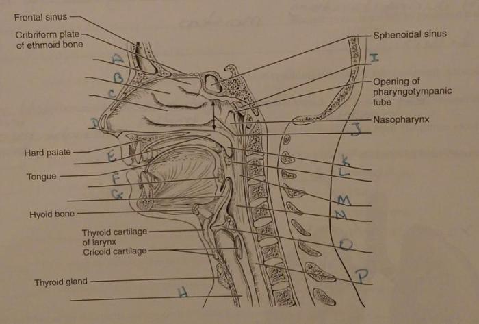
The respiratory system is a complex network of organs and tissues that allows us to breathe. It consists of the lungs, airways, and diaphragm. The lungs are two large, spongy organs located in the chest cavity. They are responsible for gas exchange, which is the process of taking in oxygen and releasing carbon dioxide.
The airways are a series of tubes that carry air to and from the lungs. They include the trachea, bronchi, and bronchioles. The diaphragm is a large muscle located at the bottom of the chest cavity. It contracts and relaxes to help us breathe.
Anatomy of the Lungs
The lungs are divided into two lobes, the right lung and the left lung. The right lung has three lobes, while the left lung has two. The lungs are made up of millions of tiny air sacs called alveoli. The alveoli are where gas exchange takes place.
They are lined with capillaries, which are tiny blood vessels. Oxygen from the air diffuses across the alveoli and into the capillaries. Carbon dioxide from the blood diffuses across the capillaries and into the alveoli.
Anatomy of the Airways, Anatomy of respiratory system exercise 36
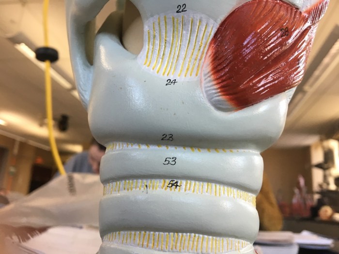
The trachea is a large tube that carries air from the nose and mouth to the lungs. It is lined with cilia, which are tiny hairs that help to move mucus and foreign particles out of the lungs. The bronchi are two large tubes that branch off from the trachea.
They carry air to the left and right lungs. The bronchioles are smaller tubes that branch off from the bronchi. They carry air to the alveoli.
Anatomy of the Diaphragm
The diaphragm is a large, dome-shaped muscle located at the bottom of the chest cavity. It separates the chest cavity from the abdominal cavity. The diaphragm contracts and relaxes to help us breathe. When the diaphragm contracts, it pulls the lungs downward, which creates a vacuum in the chest cavity.
This vacuum draws air into the lungs. When the diaphragm relaxes, the lungs recoil and air is expelled.
Respiratory System Exercise
Exercise is important for respiratory health. It helps to strengthen the lungs and improve their function. Some examples of exercises that can improve respiratory function include walking, running, swimming, and cycling.
Questions and Answers
What is the function of the diaphragm?
The diaphragm is a dome-shaped muscle that separates the thoracic cavity from the abdominal cavity. It plays a crucial role in breathing by contracting and relaxing to facilitate the inhalation and exhalation of air.
How do the cilia and mucus protect the airways?
The airways are lined with cilia, tiny hair-like structures that sweep mucus upward and outward. Mucus traps inhaled particles, preventing them from reaching the lungs and causing infection.


